")
")
Research Results 01Anomalous long-term fluorescence of rubidium vaporIn the Laboratory of Laser Physics of the Institute of Automation and Electrometry in collaboration with the Institute of Laser Physics (both of the Siberian Branch of the Russian Academy of Sciences) it discovered an interesting new phenomenon - anomalous long-term fluorescence decay time of Rb atoms in a ceresin-coated vacuum cell (see fig. 1). This time is six orders of magnitude longer than the decay time of excited rubidium atoms under normal conditions. It surprising long-term decay can only be observed in a high quality coated cell with a long lifetime of optical pumped atoms on atomic ground-state sublevels, for example, in our ceresin-coated cell with an internal atomic source. It was found that under these conditions decay is not exponential.
The study of the numerical model based on the full density matrix showed that the most likely mechanism of the observed phenomenon is the process of redistribution of population along the sublevels of the ground state of the Rb atoms. However, an understanding of the nature of the delayed decay still requires in-depth study. The discovered phenomenon is very interesting not only from the academic point of view, but can be used, for example, to study the quality of various anti-relaxation coatings in resonant cells, which is important for the creation of atomic clocks with record stability. The study was published as a preprint in the archives of Cornell University [1]. Publications
2017Spaser as a biological probe
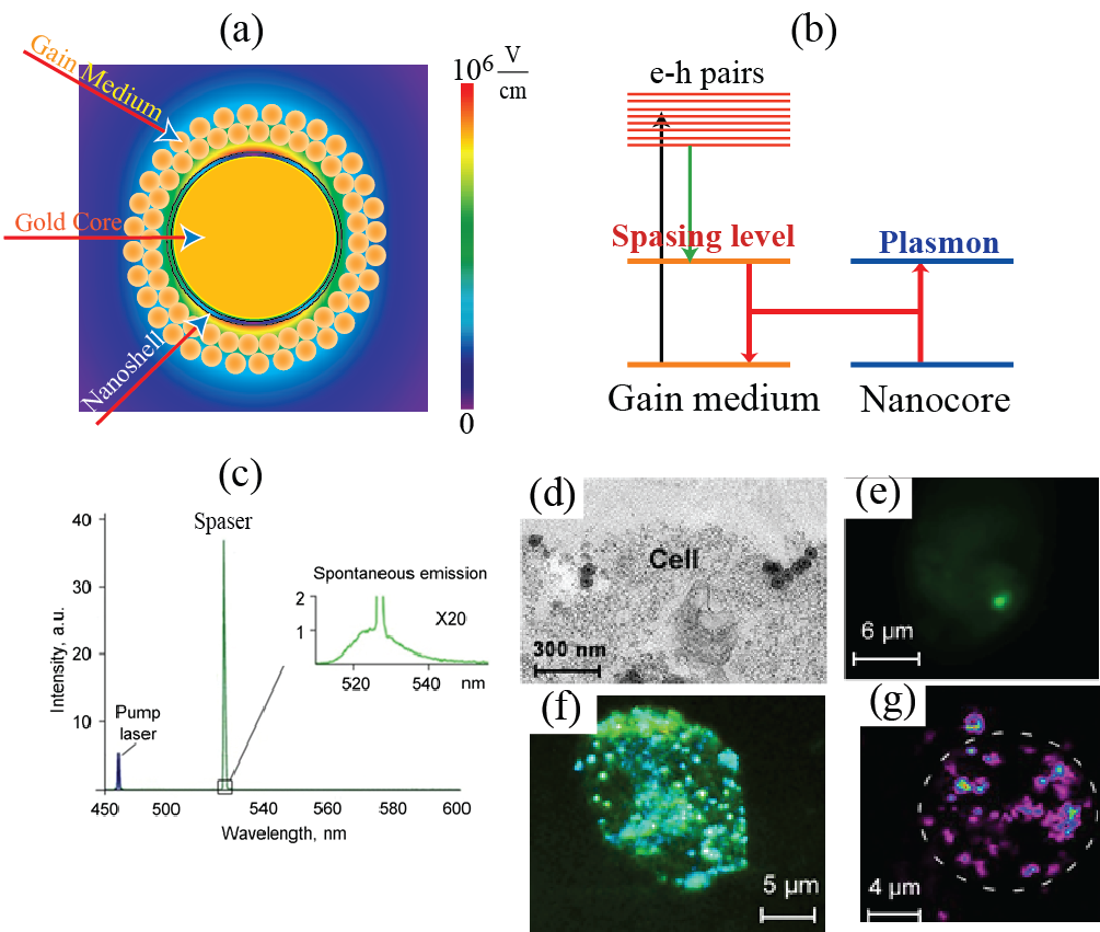
To diagnose and treat cancer, it is of primary importance to be able to see and differentiate them from normal tissues. For this purpose, one highlights the cancer cells by marking them with labels that usually are fluorescent objects: dye molecules or tiny semiconductor nanospheres called quantum dots. A requirement for such labels is that they emit bright light under optical illumination. This emission should be bright enough and have characteristic colors to be distinguishable from light scattering produced by the living tissues1. For use in vivo, these labels should be biocompatible and non-toxic for a human organism. Some of the labels can also be used for therapy, in particular, for photodynamic therapy2 or for thermal therapy3: killing the cancer cells due to photo-production of chemically active singlet oxygen or heat. Dye molecules or semiconductor colloidal quantum dots (SCQD’s) as fluorescent labels have common fundamental drawback: their emitting intensity is limited by the radiative decay rate of the fluorescing excite state (usually on the order of a nanosecond) and saturates (levels off) when the laser intensity increases to a moderate level on the order of a MW/cm2. This intensity can potentially be increased by increasing the concentration of the fluorescent label but the toxicity may become problematic in such a case, especially for the SCQD’s. Additionally, the absorption and emission lines of organic dyes are spectrally rather wide, which limits the achievable spectral selectivity. This disadvantage of wide spectra and low spectral selectivity is especially pronounced for the plasmonic nanoparticles used as theranostic agents3. A radically novel idea is pioneered in a recent article4 by Galanzha et al. where theranostic applications of a spaser (Surface Plasmon Amplification by Stimulated Emission of Radiation) are introduced and developed. The spaser as a phenomenon and device was proposed in 2003 by Bergman and Stockman5-6 and later developed and observed in a large number of works, in particular, see selected citations in ref. 4. Spaser is a plasmonic counterpart of laser where photons (quanta of electromagnetic field) are replaced by surface plasmons (quanta of electromechanical oscillations). The cavity (resonator) of a laser is replaced in the spaser by a metal nanoparticle (the spaser core), which supports surface plasmons whose fields are tightly nanolocalized in the vicinity of this core. Excitation of the spaser with an external laser pump causes generation of electron-hole pairs in the spaser shell and the stimulated near-field emission of coherent plasmons into the metal nanoparticle core (“resonator”) of the spaser – see Fig. 1 (a) and (b). Today the spasers are observed in a wide spectral range, from ultraviolet, across all visible, and into mid infrared; they are of many configuration and sizes. Now it looks like there will always be a spaser suitable for any particular nanoscopic job.
The fundamental advantage of the spaser over the conventional laser in micro- and nanoscopic application is that it is a near-field quantum generator, and its size can be much smaller than the wavelength – on the order of sizes of biological molecules and viruses. Its fundamental advantage also is that in contrast to common fluorescent labels its emission does not saturate: it is a stimulated emission and its intensity linearly depends on the pump power as long as the spaser is not physically damaged. Consequently, the brightness of the spaser and the energy released inside the targeted cell are orders of magnitude greater for the spaser than for conventional labels. The intensity and spectral brightness of the spaser radiating inside a living cell are of unprecedented total and, especially, spectral brightness – see Fig. 1 (c). A new intriguing property of transient vapor nanobubble around spaser that it can play the role of dynamic optical resonator with positive feedback amplifying spaser light and creating thus “giant” lasing that is important for cancer diagnosis at cellular levels in deep tissue. The spasers are chemically conjugated with folate to selectively target tumor cells internalized them as seen in Fig. 1 (d). The radiation of the spaser is so bright that even one spaser is well seen inside the cell [Fig. 1 (e)]. Multiple spasers produce unprecedented bright images within a cell [Fig. 1 (f)]. They are also excellent agents for photothermal and photoacoustic imaging because they also are equally efficient, unsaturable nano-generators of heat inside the cells – see [Fig. 1 (g)], which are greatly advantageous over conventional imaging and theranostics agents7-9. Moreover, ultrasharp photoacoustic resonances and red-blue resonance spitting8 can improve identification of targeted cancer cells in complex absorption background. The enormously large and unsaturable optical absorption by the spasers and the corresponding efficient generation of heat, nanobubbles8 and shock waves inside the cell allows one to effectively use them for theranostics – just a few laser pulses are sufficient to reliably kill tumor cells using photomechanical effects within the irradiated volume without damaging the healthy cells. Indeed, laser-induced nanobubbles around the heated spaser during fast expansion and collapse provide the high kinetic energy and localized pressure that can damage vital structures of triple negative breast cancer cells that are resistant to conventional therapy. One can envision that it will become possible to detect and eliminate single CTC’s as they pass through a blood vessels: a very intense and highly monochromatic (narrow-spectral-band) radiation of the spasers will allow one to continuously monitor the passage of the CTC’s through surface blood vessels. As soon as a CTC is detected via its spaser radiation, a high-power laser pulse is sent that kills these CTC’s, one tumor cell at a time. And this only the beginning of using the spaser phenomenon in biomedicine. References
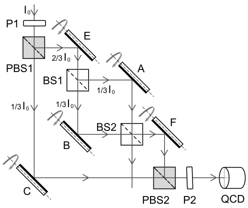
2015-2016Paradox of photons discontinuous trajectories being located by means of “weak measurements” in the nested Mach-Zehnder interferometerIn a recent letter A. Danan et al. (Phys. Rev. Lett. 111, 240402 (2013)) have experimentally demonstrated an intriguing behavior of photons in an interferometer. Simplified layout of the experimental setup is a nested Mach-Zehnder interferometer (MZI) depicted in Figure. Various mirrors inside the MZI vibrate with different frequencies. The rotation of a mirror causes a vertical shift of the light beam reflected off that mirror. The shift is measured by a quad-cell photodetector QCD. When the vibration frequency of a certain mirror appears in the power spectrum, authors conclude that photons have been near that particular mirror.
The surprising result is obtained when the inner MZI is tuned to destructive interference of the light propagating toward mirror F. In that case the power spectrum shows not only peak at the frequency of mirror C but two more peaks at the frequencies of mirrors A and B, and no peaks at the frequencies of mirrors E and F. From these results authors conclude that the past of the photons is not represented by continuous trajectories, because the photons are registered inside the inner MZI and not registered outside it. These unusual results raised a spirited discussion. Nevertheless, until now there was no comprehensive and clear analysis of the experiment within the framework of the classical electromagnetic waves approach. In particular, it was unclear if the nature of the absence of peaks at the frequencies of mirrors E and F is the same or different. We show that taking into account a) smallness of the optical beams deflection due to the mirrors vibrations, and b) axial symmetry of the beams, the light power difference absorbed by the upper (y≥0) and the lower (y≤0) cells of QCD may be represented as follows
where I0 is the light intensity at the entrance of MZI, f(x,y) is normalized amplitude distribution ( In the case of destructive interference of inner MZI (φA = φC = φB ± π) the expression in braces for difference D becomes as follows {...} = {δA - δB + δC}. Nature of the peaks absence at the vibration frequencies of mirrors E and F is different. Deflection of mirror E equally shifts beams in upper and lower arms of inner MZI, and that is why it doesn’t change its destructive interference. Deflection of mirror A (or B) results in perturbation of destructive interference of inner MZI proportional to δA (δB). Moreover, output beam from inner MZI is antisymmetrical about the y axis caused by axial symmetry of incident light beam and smallness of mirror deflection. In turn, the change of the output due to deflection of mirror F is symmetrical and is not measured, consequently, by QCD. It should be noted that measured signal D is entirely caused by the interference of modulated and unmodulated parts of light beams of MZI. So, in the case of destructive interference of inner MZI (φA = φC = φB ± π) the only unmodulated part is the one propagating along the lower arm of the outer MZI. When this light beam moving from the mirror C is blocked then all peaks disappear. So, we have clear explained paradoxical results mentioned above article, by means of traditional concept of the classical electromagnetic waves. Thus, there is no necessity for a new concept of discontinuous trajectories. Publications
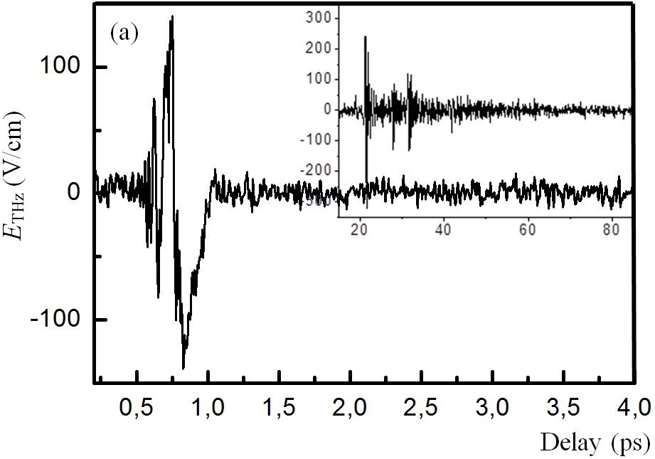
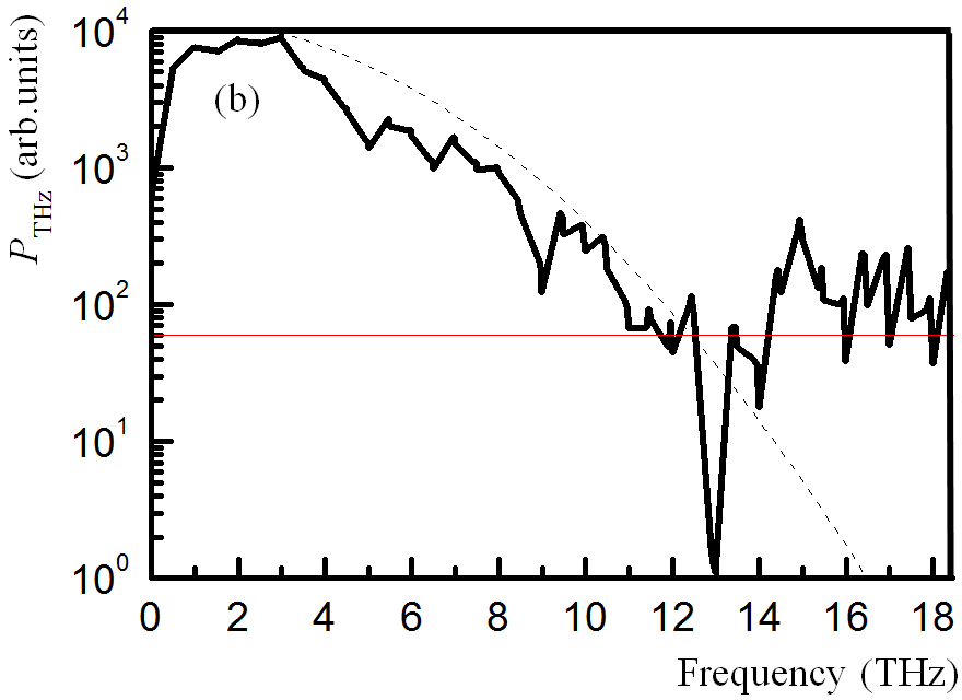
Emission of a broadband terahertz radiation in poled nonlinear optical polymersUsing the method of optical rectification of femtosecond laser pulses, we report the emission of terahertz pulses in the samples of films based on polyimides with covalently bound chromophore molecules of DR type. In sample of film which thickness is less than 1 μm a short terahertz pulses (a few field cycles, Fig. a) are excited with amplitude per unit of thickness 200 times greater than in the ZnTe crystal with a thickness of 500 μm short. The spectral width of the produced pulses is limited by the pump pulse duration (Fig. b). The quadratic nonlinear optical properties are imparted to the films in the process of their fabrication by orienting the chromophore molecules in the external electric field of the applied electrodes having an original configuration. The samples are compared with the ZnTe crystal. Using the methods of coherent spectroscopy, their transmission and refractive index dispersion spectra are investigated in the frequency range 0.5 – 2.6 THz. The polymer composition is promising for the application in coherent spectrometers both for increasing the working spectral range without dips and for improving the spatial resolution in the near-field terahertz spectroscopy.
Publications
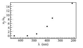
2014Photoextraction of molecular gases from a polymer organic filmWe report the first study of photodesorption of various molecular gases from a polymer organic film. This study was carried out in a Pyrex cell whose inner surface was covered by a polydimethyl-siloxane (PDMS) compound. The cell was illuminated by a flash or a CW halogen lamp. The molecular gas composition was analyzed with a mass spectrometer. We observed the variation of the molecular gas density due to photodesorption in a vapor cell as a function of illumination time, intensity, wavelength and temperature of the coating. We have observed that the desorption rate strongly depends on the light wavelength, with a threshold at about 500 nm (Figure). A linear dependence of the desorption rate on the incident light intensity has been found. This means that this effect is not caused by the direct heating of the surface and is non-thermal in nature. We have found that, under continuous illumination of the cell by a halogen lamp, the molecular photodesorption yield shows a fast decay curve, which is then followed by a long diffusion tail. The molecular photodesorption yield drops rapidly with decreasing temperature because the diffusion in the polymer in a glassy state decreases. These results are a clear indication that bulk diffusion plays an important role in the observed molecular photodesorption process. This study could be useful for constructing light-driven sources of molecules.
Publications
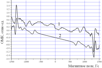
2013Narrow dark resonances in the spontaneous emission of gas mixture of the even isotopes of neonThe new phenomenon – the suppression of spontaneous emission – has been observed experimentally in a gas mixture of isotopes 20Ne and 22Ne. The effect is appeared as the narrow optomagnetic resonances (OMRs) in changing of intensity of the glow of the gas mixture which was in a scanned longitudinal magnetic field. The positions of OMRs (±1400 Gs and ± 900 Gs) are defined by the resonance conditions, when the isotope shift is compensated by the Zeeman effect. The narrowness of the widths of OMRs shows diminishing of the Doppler effect, i.e. atoms of different isotopes that contribute into the resonances are in rest to each other. In these conditions there is a correlated spontaneous emission of a pair of isotopes with a reduced probability.
Isotopic OMRs are observed at low gas pressure p ≈ 0.2 mm Hg and at narrow range of pressure – Δp/p ~ 0.1. To create the OMRs the presence of both isotopes –20Ne and 22Ne are required. Resonances at ±1400 Gauss correspond to decreasing of light emission of the gas mixture. The obtained results of the experiment are in accordance with hypothesis of formation of a pair of entangled states of atoms of different isotopes of neon. Publications
Light desorption of molecular nitrogen from a glass surface
Publications
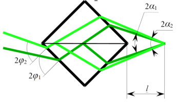
Two-ray interferometers tuned by rotation of the device itself
Publications
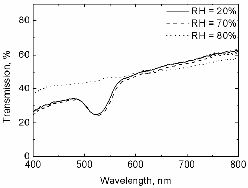
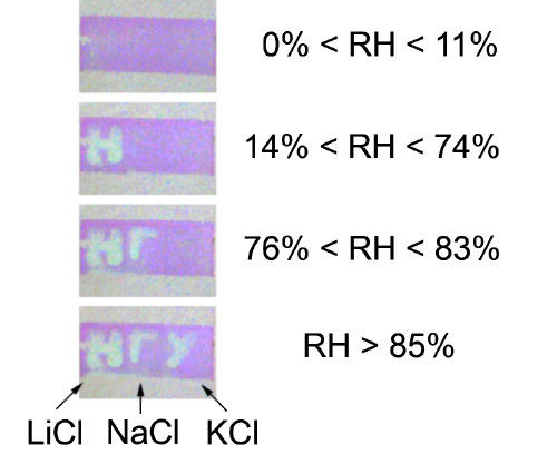
Optical humidity sensor based on photonic crystal opal filmA new type of colorimetric sensor for humidity measurements has been proposed on the basis of photonic crystal (PhC) opal films. Sensor does not require electricity and represent a PhC plate saturated by salt solutions on different areas, which becomes transparent with increasing of humidity more than a defined value. The discovered effect is caused by vanishing of the band gap in transmission spectrum of the PhC film (Fig. 1). Upon reduction of humidity, the film regain its initial spectral properties in tens seconds. The revealed effect gives an opportunity to detect more than 50 levels of humidity, which is determined by selection of slats. The sensor has high temporal stability and allows registering a relative humidity change with precision of 2%.
Publications:
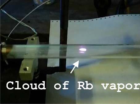
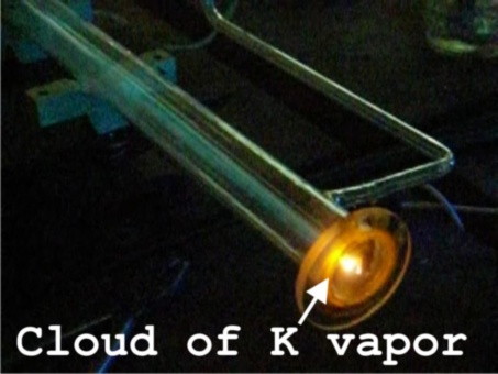
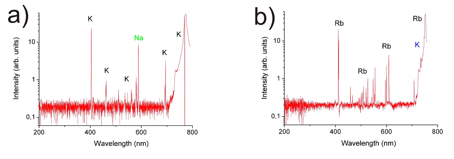
2012Explosive evaporation of Rb or K fractal clusters by low power CW radiation in the presence of excited atomsWe describe a new, spectacular, unpredictable effect of the explosive evaporation of metallic Rb or K fractal clusters, only in the presence of excited atoms stimulated by resonant CW laser radiation in a heat - pipe glass cell (Fig.3a,b, see also the next moves Potassium.avi, Potassium on the window.avi, Rubidium.avi).
Evaporation occurs at low laser-power density, in the presence of a buffer gas. The effect consists of the generation of optically thick, sharply localized alkaline metals vapour clouds propagating in the cell against the laser beam. These clouds are charged and exhibit a strong luminescence of Rb or K spectral lines.
We believe that the explosive evaporation of metallic fractal clusters observed is explained by the laser excitation of alkali atoms. The excited atom collides into the surface of the clusters and transfers its internal energy to the surface locally. This energy greatly raises the temperature of this local part of the clusters surface, melts it and decreases the fractal surface area. Because, in general, any fractal cluster systems have a high surface energy, any process which leads to decreasing their surface area can liberate the surface energy. This energy increases the total temperature of the clusters and eventually leads to the thermal explosion of the cluster. Publications
|
||||||||||||||||||||||||||||||||||
|
Institute of Automation and Electrometry of the Siberian Branch of the Russian Academy of Sciences |
 |
-
Home

-
Labs Pages
-
Laboratory 01
- Research Results 01
 Cancer tumors are known for their ability to proliferate or metastasize. This happens either through cancer cells spreading through lymphatic nodes or by the so called circulating tumor cells (CTC’s). In the difficult task of cancer diagnostics and therapeutics (the so-called theranostics), the easiest task may be removal or destruction of the primary tumor but fighting the cancer cells migrating through the lymphatic system at the CTC’s traveling through blood vessels is an altogether different and much more difficult problem.